Image: Carolin Lerch / TUM
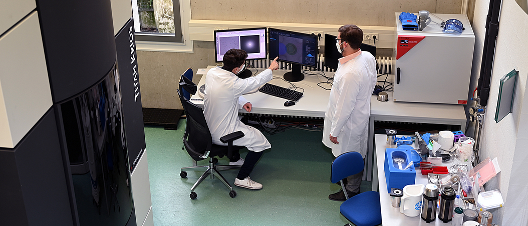
At MIBE, two 300-kV transmission electron microscopes (TEMs) enable imaging with sub-nanometer resolution.
In both TEMs (Thermo Fisher Titan Krios and JEOL JEM3200 FSC), highly coherent electrons extracted from field emission sources are accelerated to 300 keV in a high voltage potential. The electron beam is focused using electromagnetic lenses. When shining through the sample, the electrons are scattered and the resulting image which is then captured by a detector corresponds to a transmission projection of the sample.
With the help of a direct electron detector, single incident electrons can be detected to achieve a high signal-to-noise ratio and thus high-resolution images, which is crucial in particular with dose-sensitive samples. Special technical equipment consisting of an aberration corrector as well as a contrast amplifying phase plate and energy filter are used to increase the quality of the images.
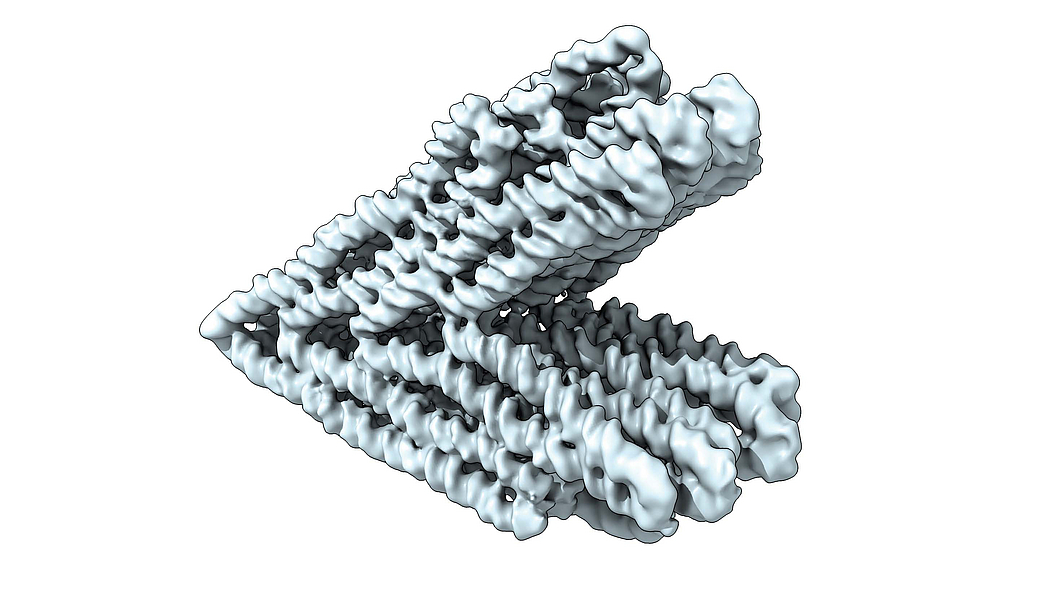
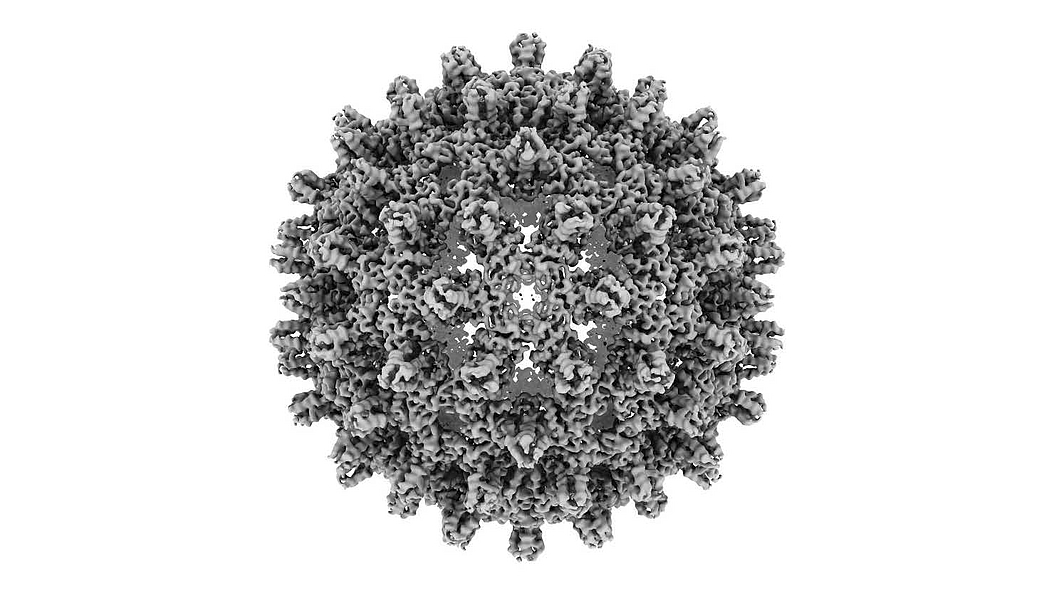
In addition to material science research, a special focus lies in imaging biological samples such as proteins and DNA structures at cryogenic temperatures. These images can then be used to reconstruct their three-dimensional structure to provide valuable information about shape and function.
As part of the TUM Electron Microscopy Core Facility of the Competence Area Bio- and Materials Sciences, the TEMs are currently operated and managed by the research group led by Prof. Hendrik Dietz.
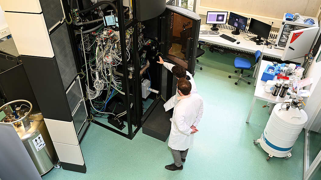
Image: Carolin Lerch / TUM
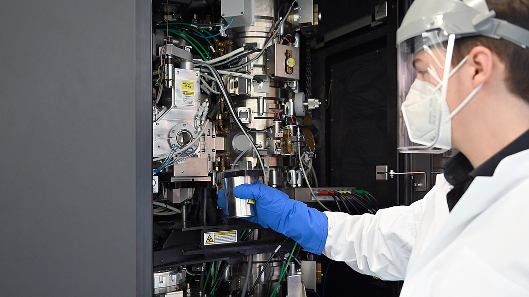
Image: Carolin Lerch / TUM
