Medizinische Bildgebung & Strahlentherapie
Im Bereich der medizinischen Bildgebung und Strahlentherapie beschäftigen sich Forschende am MIBE mit neuartigen Technologien für Computertomographie oder Magnetresonanzbildgebung, erforschen die radiobiologische Wirkung von neuartigen Strahlentherapiekonzepten, oder entwickeln neuartige Tracer und Kontrastmittel mit spezifischer molekularer Sensitivität.
Phasenkontrast Röntgen-Bildgebung
PI Franz Pfeiffer
Lehrstuhl für Biomedizinische Physik
Publikationen
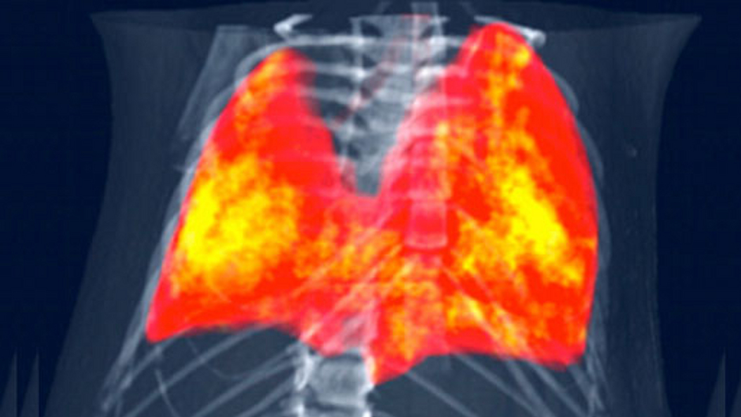
Konventionelle Röntgenbildgebung nutzt nur einen Teil der in den Röntgenstrahlen enthaltenen Information. Die Phasenkontrast-Methode macht mehr Information zugänglich und erzeugt damit Bilder, in denen viele unterschiedliche Gewebetypen deutlich voneinander unterschieden werden können. Die Methode ist in den letzten Jahren am Lehrstuhl von Prof. Franz Pfeiffer soweit entwickelt worden, dass sie demnächst routinemäßig in der klinischen Diagnostik zum Einsatz kommen kann.
Physik der Biomedizinischen Bildgebung
PI Julia Herzen
Forschungsgruppe Physik der Biomedizinischen Bildgebung
Publikationen
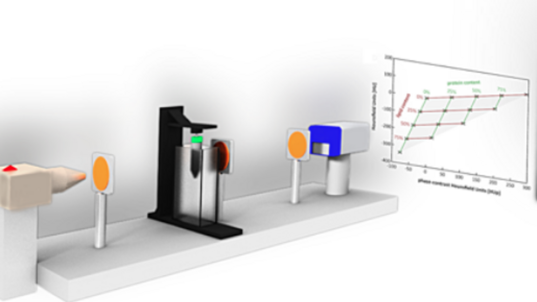
The team around Prof. Herzen develops novel X-ray imaging methods using highly brilliant synchrotron radiation and conventional laboratory X-ray sources. They mainly focus on quantitative multi-modal approaches combining spectral and phase-contrast imaging. Currently, they are aiming at applying these methods for improved breast cancer detection and for quantitative 3D virtual histology of human tissue.
Weitere Informationen:
TEDx Talk (Youtube-Video): Novel X-Ray technology that can revolutionize preventive medicine
NMR-Bildgebung
PI Axel Haase
Munich Institute of Biomedical Engineering
Publikationen
Nuclear Magnetic Resonance (NMR) is a spectroscopic method, which is commonly used in science to elucidate the structure and dynamics of molecules. It uses strong magnetic fields (up to 23 Tesla) with highest homogeneity, temporal stability, and with radio frequency fields (up to 1 GHz). The additional use of magnetic field gradients (up to 1 Tesla/m) allows for the acquisition of MR images.
In general, Magnetic Resonance Imaging (MRI) uses the 1H MR signal from water and lipid molecules to create 2D or 3D images of biological objects (organs, animals, plants, human subjects). MRI is a non-invasive method and shows no biological side effects.
Therefore it is ideally suited to study and visualize the anatomy, function and metabolism of internal organs.
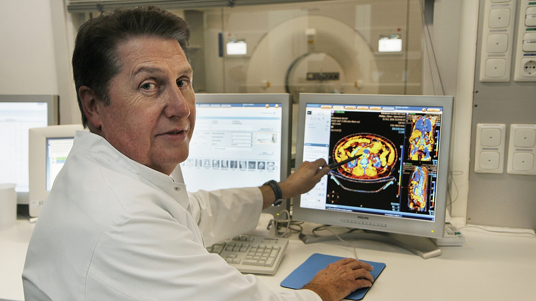
Theranostics
PI Wolfgang Weber
Professur Nuklearmedizin
Publikationen
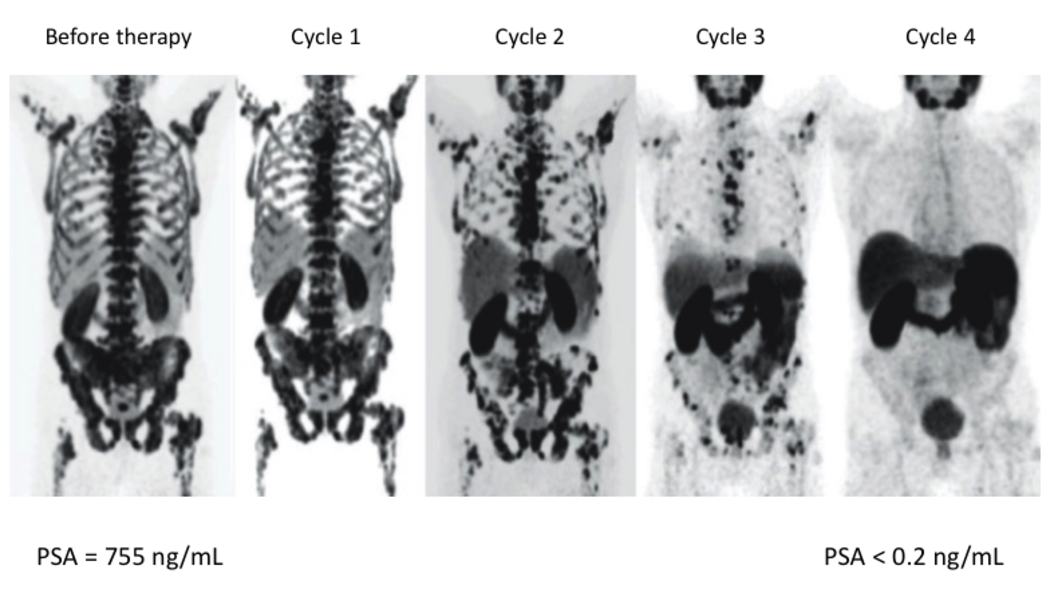
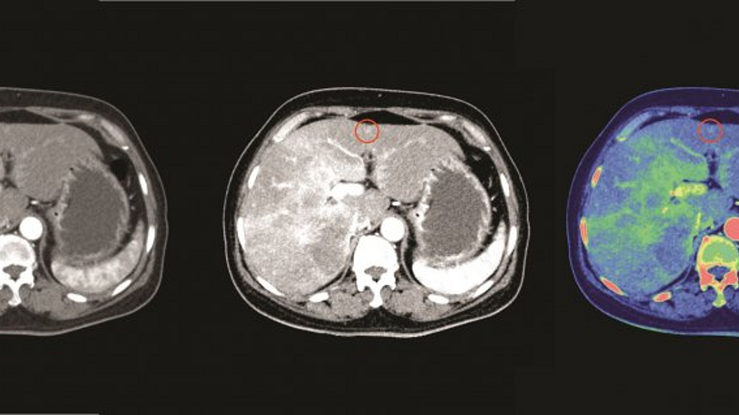
Theranostics, the combination of imaging and targeted radionuclide therapy, aims to treat cancer by radioisotopes that specifically accumulate in the tumor tissue due to molecular targeting. Theranostics expands the use of radiotherapy - one of the most effect therapies for virtually all cancers - to the treatment of patients with metastatic disease. Quantitative nuclear imaging is used to select patients for targeted radionuclide therapy, calculate radiation doses and monitor tumor response.
Biologische & Optische Bildgebung
PI Vasilis Ntziachristos
Lehrstuhl für Biologische Bildgebung
Publikationen
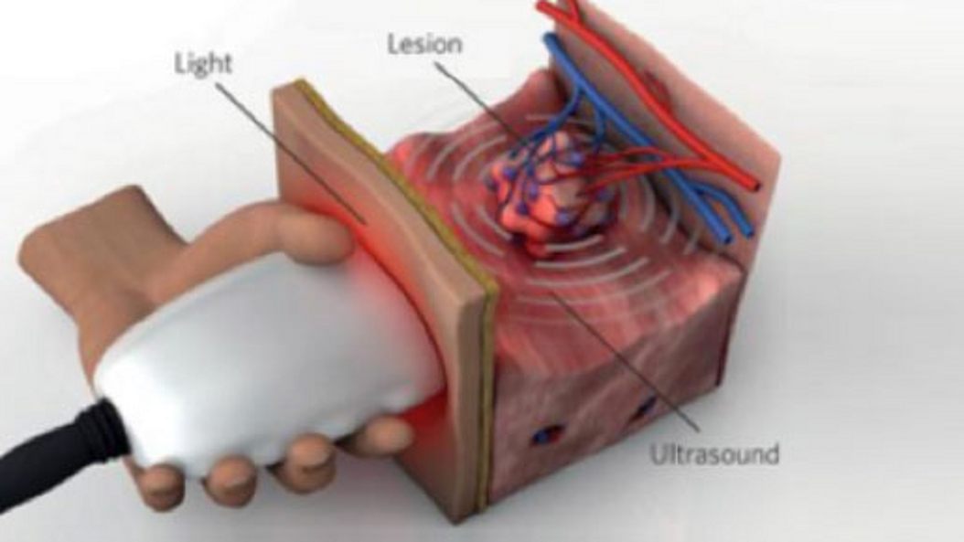
Optoacoustic imaging, or photoacoustic imaging, is insensitive to photon scattering within biological tissue and, unlike conventional optical imaging methods, and makes high-resolution optical visualization deep within tissue possible. Recent advances in laser technology, detection strategies and inversion techniques have led to significant improvements in the capabilities of optoacoustic systems. A key empowering feature - pioneered at TUM - is the development of video-rate multispectral imaging in two and three dimensions, which offers fast, spectral differentiation of distinct photo-absorbing modalities.
Experimentelle Magnetresonanztomographie
PI Dimitrios Karampinos
Forschungsgruppe Experimentelle Magnetresonanztomographie
Publikationen
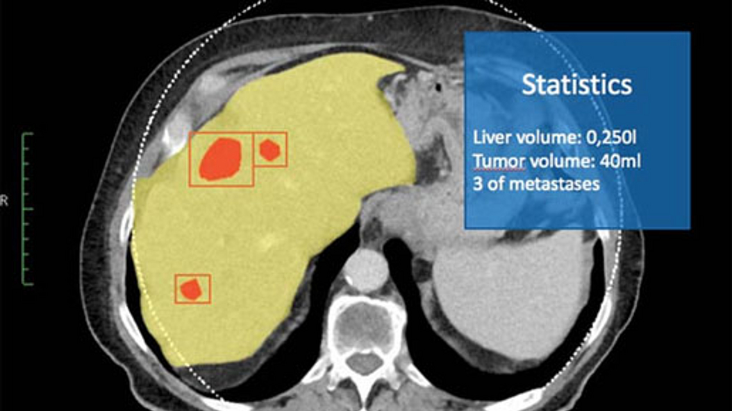
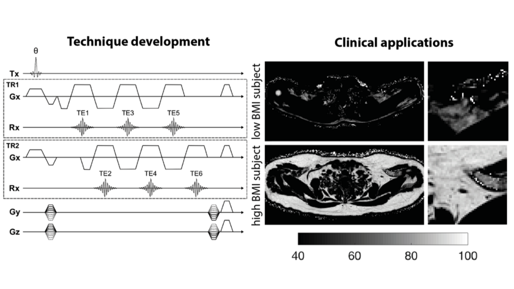
Prof. Karampinos' research focuses on the development of novel Magnetic Resonance Imaging (MRI) and Magnetic Resonance Spectroscopy (MRS) acquisition, reconstruction and signal modeling methods with an emphasis on the extraction of quantitative imaging biomarkers. He and his team are developing novel methods for proton density fat fraction mapping, lipid diffusion mapping, quantitative susceptibility mapping, T2 and T2* mapping, diffusion tensor imaging and body diffusion-weighted imaging. The developed methods are being translated into clinical studies for improving the diagnosis, the therapy monitoring, and the understanding of disease pathophysiology in the diseases of the musculoskeletal system (e.g. osteoporosis, neuropathies, neuromuscular diseases), in metabolic dysfunction (e.g. obesity, diabetes, cachexia) and in body oncology.
Magnetic Resonance Imaging Biomarkers
PI Franz Schilling
Forschungsgruppe Biomedizinische Magnetresonanz
Publikationen
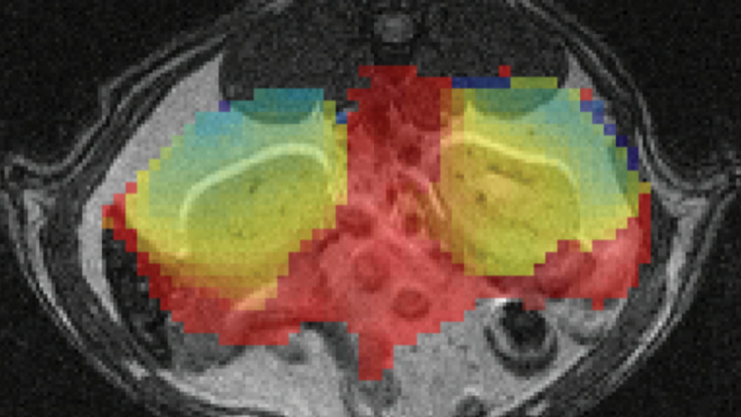
Magnetic resonance imaging biomarkers enable a comprehensive characterization of tissue providing functional, physiological, metabolic, cellular and molecular information beyond anatomical structures. In recent years, hyperpolarization techniques allowed to increase the inherent low sensitivity of magnetic resonance by more than four orders of magnitude, opening new applications ranging from the tracking of chemical reactions in real-time to the direct monitoring of metabolic processes in vivo. Another functional MRI technique that has already shown promise in characterizing tissue in clinical studies is diffusion-weighted MRI (DW-MRI). It enables the quantitative assessment of the apparent diffusion coefficient (ADC) that depends on microstructural tissue properties, including cellularity and intercellular matrix composition. At TUM, sensitive hyperpolarized molecules and advanced diffusion MRI techniques are being developed aiming to provide novel imaging biomarkers to characterize tissue function.
X-Ray & Computed Tomography
Prof. Dr. Daniela Pfeiffer
Professur für Diagnostic & Interventional Radiology
Publikationen
Daniela Pfeiffer is an expert in the field of lung imaging and musculoskeletal diagnostics. Her main medical research interest is improving detection, precise diagnosis and therapy follow-up of lung diseases such as COPD, lung cancer and infectious diseases (including COVID-19).
From a technical perspective, she is mainly interested in novel methods for medical X-Ray and Computed Tomography (CT) Imaging. Her main focus lies in the field of photon counting CT technology, spectral CT applications, dark-field and phase contrast imaging, and machine learning in clinical applications.
Diagnostische Radiologie
Prof. Dr. Ernst Rummeny
Professur für Radiologie
Publikationen
Prof. Rummeny war Direktor des Instituts für Radiologie und Arzt für Diagnostische Radiologie und Nuklearmedizin. Seine Forschungsschwerpunkte lagen im Bereich der Onkologischen Bildgebung, insbesondere in der Entwicklung der Schnittbildverfahren wie MRT- und CT-Tomographie und damit verbundener Hybridsysteme, wie PET/CT und MR-PET zur Erkennung und Therapiebeurteilung von malignen und benignen Erkrankungen. Hierfür entwickelte er zusammen mit Kollegen der Biomedizinische Physik (Prof. F. Pfeffer) und Kollegen der Biologischen Bildgebung (Prof. V. Ntziachristos) neuartige Geräte, die translational für erste Untersuchungen an Patienten eingesetzt wurden. Darüber hinaus war er an der Optimierung von Kontrastmitteln beteiligt.
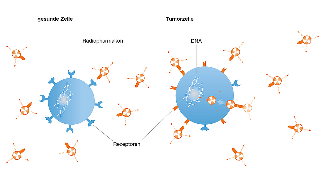
Pharmazeutische Radiochemie
Prof. Dr. Hans-Jürgen Wester
Lehrstuhl für Pharmazeutische Radiochemie
Publikationen
Die Forschung von Herrn Prof. Wester konzentriert sich auf die Entwicklung gezielt radioaktiver markierter Proben und Therapeutika (Radiopharmakon) zur molekularen Bildgebung sowie gezielter Behandlung mit Radioliganden. Er entwickelt mehrere Verfahrensweisen und Radiopharmakons für onkologische, neurologische und cardiovaskuläre Untersuchungen und Therapien, die eine breite Anwendung in klinischen Untersuchungen und Therapien gefunden haben.