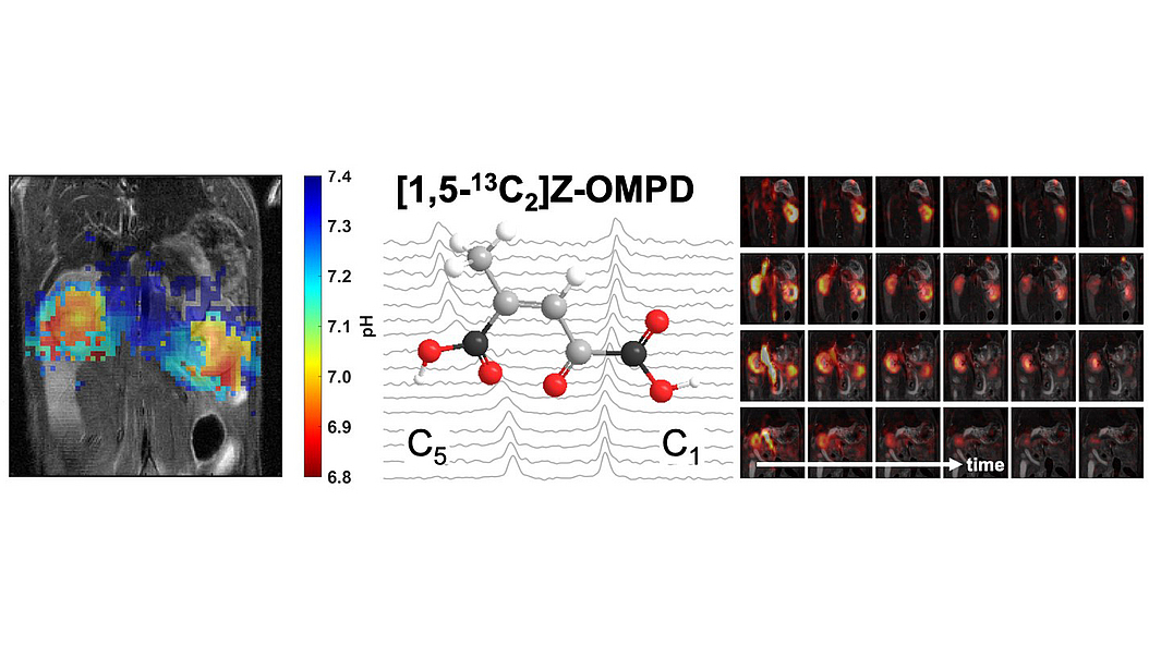
Visualizing the pH value in the human body could facilitate earlier and more precise diagnosis of kidney disease and cancer, and speed the assessment of a therapy's efficacy. Until now, however, there has been no non-invasive way to measure this value in everyday clinical practice. Researchers at the Technical University of Munich (TUM) have identified a molecule for magnetic resonance imaging (MRI) which can represent the acid to base ratio in kidneys and tumors.The molecule occurs naturally in tulips. According to initial findings, it has significant advantages over other substances investigated to date.
"The aggressiveness of tumors, or in other words, the potential of a tumor to spread and further attack the body, often correlates closely with a change in metabolism. Tissue can become highly acidic in the vicinity of tumor cells. This helps the tumor overcome healthy cells and continue growing," says Franz Schilling, Professor of Biomedical Magnetic Resonance at TUM. Together with his team he is working on molecules that enable imaging of the pH value in the body in order to render such metabolic effects visible and make them measurable for viable use in diagnostics.
The researchers have now identified a molecule for MRI called Z-OMPD, which they can synthesize in the laboratory. In preclinical tests with mice and rats this molecule displays particularly useful properties for imaging. In order to make it visible in MRI, it is marked at two places with carbon-13. In contrast to carbon-12, which is significantly more common, carbon-13 is magnetic and generates a signal in MRI. Since this signal is relatively weak, it is amplified in a process known as hyperpolarization. Carbon-13 is not radioactive and does not expose the patient to any radioactivity.
Molecule occurs in tulips
"Just recently the parallel discovery was made that Z-OMPD occurs naturally in tulips in high concentrations. In addition, it's assumed that this substance also occurs in exobiological processes on interstellar asteroids and is thus already fundamentally biocompatible," says Martin Grashei, one of the study's two lead authors.
"One of the major advantages of this molecule for imaging is its longer magnetic half-life compared to many other hyperpolarized molecules. This means we see the signal in the magnetic field for a longer period of time, which lets us achieve higher image quality," says Pascal Wodtke, the other lead author of the study.
Advantages in imaging of the kidneys
In addition to its use in oncology, Z-OMPD could also be particularly well suited for renal imaging. Our kidneys filter approximately 1500 liters of blood every day, removing substances which are harmful or no longer needed by the body. In case of kidney disease the pH value in the renal system can be impaired. Z-OMPD could be used to render this visible at an early stage in an MRI examination, followed by a more in-depth investigation of the affected areas.
Another factor which is especially favorable for use in the kidney is the fact that Z-OMPD also enables non-invasive measurement and quantitative evaluation of tissue perfusion and the renal filtration rate in one and the same measurement. This provides additional information for the more precise delineation of the clinical picture. As yet, there is no non-invasive method for measuring the pH value in use in everyday clinical practice. Perfusion and filtration are often measured using radioactively marked substances or contrast agents containing gadolinium, both of which often entail complications and risks for the patient.
Measuring the filtration rate using MRI imaging has the additional advantage that the individual kidneys can be specifically examined. Filtration measurement using for example blood values on the other hand can only provide information about both kidneys collectively.
Stable measurements
To investigate the pH value, molecules marked for imaging are injected into the tissue, where they serve as sensors. The nuclear magnetic resonance frequency of the molecules varies depending on the pH value. Researchers can measure this frequency value and use it to determine the pH value.
The frequency can however also shift due to other effects such as fluctuations in the MRI magnetic field. The principle resembles that of a violin which is out of tune and has to be corrected using a tuning device. Accordingly the researchers usually inject a second substance as a reference which remains stable compared to the changes in the pH value. This makes it possible to calculate and correct for undesired effects. "That's not necessary with Z-OMPD. The molecule has an internal reference and already contains a second signal which remains stable during injection," says Franz Schilling.
The advantages of imaging with Z-OMPD have already been demonstrated with mice and rats. The researchers are now working to further improve the method so that it can be tested in clinical studies. This new technique to image the pH value could help make earlier and more precise diagnoses possible. Cases of suspected kidney problems could be resolved faster, and transplanted kidneys could be monitored after implantation. The method could also help in better predicting the success of tumor therapies before treatment and could speed assessment of the therapies afterwards.
Scientific graphical abstract

Publication
Martin Grashei, Pascal Wodtke, Jason G. Skinner, Sandra Sühnel, Nadine Setzer, Thomas Metzler, Sebastian Gulde, Mihyun Park, Daniela Witt, Hermine Mohr, Christian Hundshammer, Nicole Strittmatter, Natalia S. Pellegata, Katja Steiger, Franz Schilling: Simultaneous Magnetic Resonance Imaging of pH, Perfusion and Renal Filtration using Hyperpolarized 13C-labelled Z-OMPD, Nature Communications (2023), DOI: doi.org/10.1038/s41467-023-40747-3
More Information
Prof. Franz Schilling conducts research at the Klinikum rechts der Isar of TUM and at the Munich Institute of Biomedical Engineering (MIBE), an Integrative Research Institute within TUM. At MIBE, researchers specializing in medicine, the natural sciences, engineering, and computer science join forces to develop new methods for preventing, diagnosing or treating diseases. The activities cover the entire development process – from the study of basic scientific principles through to their application in new medical devices, medicines and software.
The project was funded by the German Research Foundation (DFG) as part of the Collaborative Research Center SFB824, by the Young Academy of the Bavarian Academy of Sciences and Humanities, by the European Union's Horizon 2020 research and innovation program, and by the program "Quantum Technologies - from Basic Research to Market" / "QuE-MRT" of the German Federal Ministry of Education and Research (BMBF).
Contact Media Relations
Corporate Communications Center
Technical University of Munich
Carolin Lerch
carolin.lerch@tum.de
Media Relations MIBE
presse@bioengineering.tum.de
Scientific Contact
Prof. Dr. Franz Schilling
Technical University of Munich
Professor of Biomedical Magnetic Resonance
Tel: +49 89 4140 4586
fschilling@tum.de
