News Biomedizinische Mikroskopie & Virtuelle Pathologie

  • Julia Herzen, Professorin für Biomedizinische Bildgebung

    Hochaufgelöste Röntgentechnik weiterentwickeln

    ERC Consolidator Grant für Julia Herzen

    23. November 2023 | Prof. Julia Herzen erhält einen ERC Consolidator Grant für das Projekt DEPICT. Sie entwickelt ein physikalisches Modell, um hochauflösende Röntgenbildgebung auf der Mikrometerskala entscheidend weiterzuentwickeln und damit zukünftig zum Beispiel Zusammensetzung, Verteilung und die Menge einzelner Materialien bestimmen zu können.


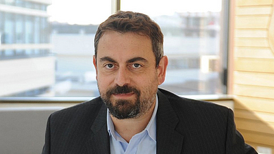
  • Vasilis Ntziachristos, Professor für Biologische Bildgebung hat mit seinem Team die optoakustische Bildgebungsmethode RSOM entwickelt. Bild: Stefan Rumpf

    Innovationspreis der Europäischen Kommission für INNODERM

    Tiefe Einblicke mit dem Haut-Scanner

    15. Dezember 2021 | Innovationspreis der Europäischen Kommission für INNODERM

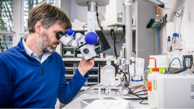
  • Prof. Thomas Misgeld ist Direktor des Instituts für Zellbiologie des Nervensystems an der TUM und Koordinator des Exzellenzclusters SyNergy. Bild: ediundsepp

    Entzündung verursacht Fehlfunktion der Hirnrinde bei Multipler Sklerose

    Immunzellen greifen Synapsen an

    27. Januar 2021 | Schädigungen der grauen Hirnsubstanz tragen maßgeblich zum Fortschreiten der Multiplen Sklerose bei. Ein Team von Neurowissenschaftlerinnen und -wissenschaftlern hat jetzt gezeigt, dass die Ursache Entzündungsreaktionen sind, die zu Synapsenverlust führen und so die Aktivität der Nervenzellen vermindern. 

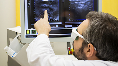
  • Prof. Vasilis Ntziachristos demonstriert eine der multi-spektralen optoakustischen Mesoskopie ähnliche Bildgebungsmethode. (Bild: M. Jooss)

    Optoakustische Mesoskopie visualisiert Gewebemuster von Tumoren

    Hochauflösende 3D-Ansicht des Tumorinneren

    27. Mai 2020 | Einem Forschungs-Team der TUM und des Helmholtz Zentrums München ist es mit Hilfe der optoakustischen Bildgebung gelungen, räumliche Veränderungen von Tumoren sichtbar zu machen. 

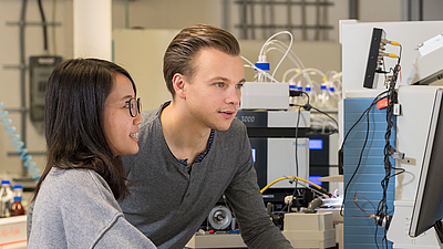
  • Researchers at the Chair of Proteomics and Bioanalytics (Prof. Dr. Bernhard Kuester) at the TUM School of Life Sciences Weihenstephan (WZW). Image: Astrid Eckert / TUM

    CLINSPECT-M: Clinical mass spectrometry center for molecular brain research

    Major project for Munich neurosciences

    10 Feb 2020  | In a joint large-scale project, Munich scientists from proteomics, computer science and medicine investigate the causes of disorders of the central nervous system, how they can be diagnosed and how treatment response can be monitored.

  • Distribution of a drug in the lung of a mouse at two different times after instillation.

    X-rays and fluorescence imaging of the lungs of a mouse enable tracking of drug delivery

    Precise monitoring of the lung

    14 Jan 2020 | A combination of imaging techniques allows real-time monitoring of the delivery of inhaled drugs within the lungs of mice and their absorption in the lung tissue.

  • MiROM micrograph of living adipocytes

    Looking at the good vibes of molecules

    A new method for label-free metabolic imaging

    09 Jan 2020 | A novel imaging method will have a major impact on basic understanding, monitoring, prevention, diagnosis, and treatment of metabolic diseases, such as diabetes and obesity.

  • MSB-PI Vasilis Ntziachristos

    Radio feature about optoacoustic tomography (in German)

    04 September 2019 | German public radio (Deutschlandfunk) reported on the work of MSB-PI Vasilis Ntziachristos and his colleagues at the TUM professorship of Biological Imaging and the Institute for Biological and Medical Imaging at the Helmholtz Zentrum München.