Contrast agents are often used to improve the imaging of soft tissue in micro-computed tomography (microCT). Now a research team led by the Technical University of Munich (TUM) has investigated how these agents disperse inside cells. Their findings could improve the assessment and further development of contrast agents and might contribute to future medical diagnostics.
Contrast agents are used very frequently in the difficult task of rendering organ structures visible using conventional X-rays in microCT. The question of the exact concentrations and final locations of these agents within cells has in the past remained largely unexamined, since doing so requires high-resolution methods which are also extremely sensitive.
Now a research team led by Julia Herzen, Professor of Biomedical Imaging Physics at TUM has addressed this question for two previously known exemplary contrast agents. The first, iodine potassium iodide, stains cells unspecifically, which means it does not interact with specific structures. The second contrast agent studied, Eosin Y, contains bromine and dyes the cell cytoplasm, the interior of the cell, since it bonds to protein structures found there.
Cells with contrast agents examined at particularly high resolutions
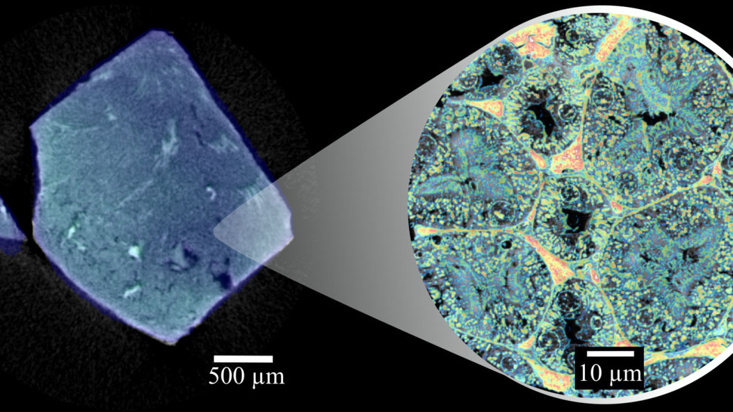
After dying with the contrast agents, the tissue samples were first analyzed using dual-energy microCT (MicroDECT), a quantitative X-ray imaging method. The researchers used the procedure to determine the overall concentrations of the contrast agents in the tissue. However, this method's resolution is not high enough to represent individual cells.
The next step was to determine how much of the contrast agent had accumulated within the cells and to which individual structures. Here the researchers used a highly sensitive and high-resolution method referred to as ptychographic X-ray computed tomography (PXCT) in a facility at Switzerland's Paul Scherrer Institute. This method is suitable for quantitative analysis and can represent soft tissue as well as organs in greater detail than MicroDECT can.
Using MicroDECT, both contrast agents provided images with comparable levels of contrast. It took ptychography to reveal the significantly different accumulation of the two contrast agents in the cells. “It was surprising to find that for both contrast agents, only very low concentrations were measured in the cells compared to the respective initial solution we used for staining“, says first author Kirsten Taphorn. "In general, we found that Eosin Y accumulated significantly less in the cells than the iodine-based contrast agent."
Potential further development for diagnostic applications
In the current study the research team investigated two previously known contrast agents. "In the future, ptychography could be used to analyze newly developed contrast agents and to make sure that they actually bond with the desired structures. This would be of particular interest for contrast agents which bond with very specific structures, for example with a certain protein in tumor tissues. This new contrast agent could then be used in combination with virtual X-ray based histology," says Prof. Julia Herzen, head of the study.
Publication
Madleen Busse, Johannes Brantl, Benedikt Günther, Ana Diaz, Mirko Holler, Martin Dierolf, Doris Mayr, Franz Pfeiffer, Julia Herzen: X-ray Stain Localization with Near-Field Ptychographic Computed Tomography. Advanced Science, June 2022. DOI: https://doi.org/10.1002/advs.202201723
More Information
The authors Prof. Julia Herzen, Prof. Franz Pfeiffer, Kirsten Taphorn, Dr. Madleen Busse, Johannes Brantl, Dr. Benedikt Günther und Dr. Martin Dierolf conduct research at the Munich Institute of Biomedical Engineering (MIBE). MIBE is an Integrative Research Institute (IRI) within the Technical University of Munich (TUM) that fosters interdisciplinary cooperation and synergies between researchers from the broad field of Biomedical Engineering. At MIBE, researchers specializing in medicine, the natural sciences, and engineering join forces to develop new methods for preventing, diagnosing or treating diseases. The activities cover the entire development process – from the study of basic scientific principles through to their application in new medical devices, medicines and software.
Researchers from the following institutions contributed to the study: Technical University of Munich (TUM), Paul Scherrer Institut (PSI), Ludwig-Maximilians-Universität Munich (LMU).
Contact Media Relations
Contact MIBE Media Relations:
presse@bioengineering.tum.de
Scientific Contact
Prof. Dr. Julia Herzen
Technical University of Munich
Professor of Biomedical Imaging Physics
Phone: +49 89 289-10806
Julia.herzen@tum.de


