Biomedical Microscopy & Virtual Pathology
In Biomedical Microscopy and Virtual Pathology, MIBE scientists explore novel technological concepts for mesoscale and microscopic three-dimensional biological imaging to better understand the causes and progression of diseases.
Biological & Optical Imaging
PI Vasilis Ntziachristos
Chair of Biological Imaging
Publications
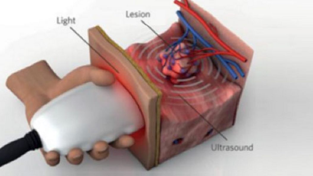
Optoacoustic imaging, or photoacoustic imaging, is insensitive to photon scattering within biological tissue and, unlike conventional optical imaging methods, and makes high-resolution optical visualization deep within tissue possible. Recent advances in laser technology, detection strategies and inversion techniques have led to significant improvements in the capabilities of optoacoustic systems. A key empowering feature - pioneered at TUM - is the development of video-rate multispectral imaging in two and three dimensions, which offers fast, spectral differentiation of distinct photo-absorbing modalities.
Morphomolecular Imaging
PI Wilko Weichert
Institute of General & Surgical Pathology
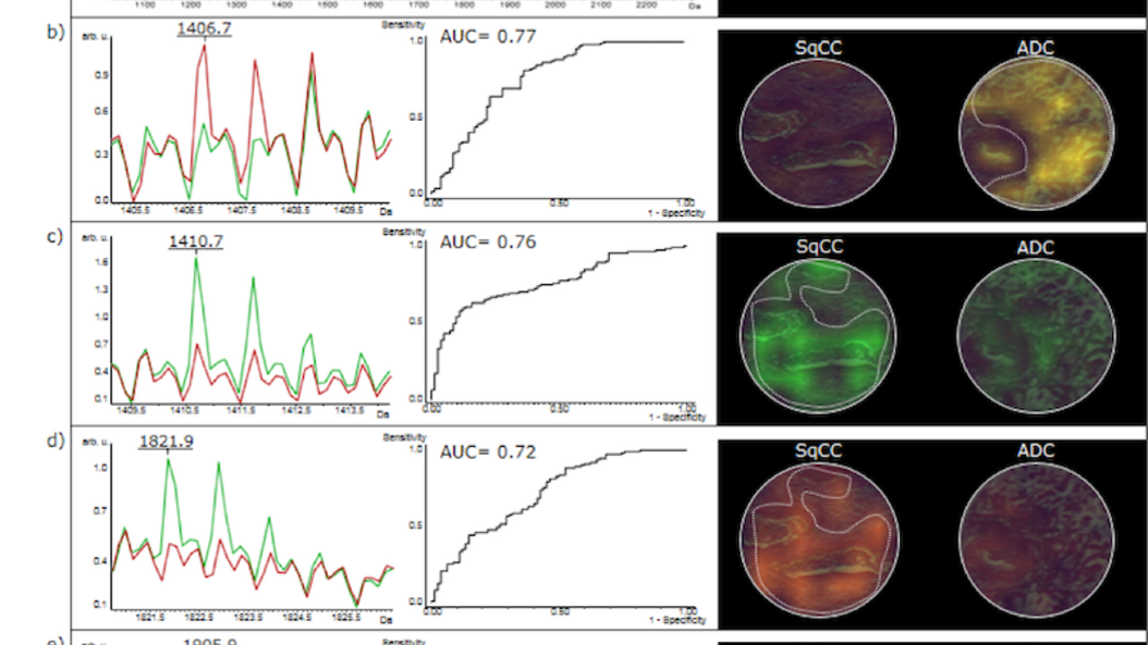
Tissue based morphomolecular “imaging” and deep multiparameter characterization has become one of the cornerstone of individualized patient care. The integration of histology with molecular information such as sequencing results or imaging mass spectrometry data allows for a deeper biological understanding of diseases and thus for tailored therapeutic approaches. This is specifically important in the field of oncology where such integrated multiparameter “imaging” datasets already dictate therapeutic decisions and allow for the application of novel drugs which prolong patient survival substantially. To exploit the tremendous new set of morphomolecular disease information at hand new methods of bioinformatics and computer learning have to be applied and will further deepen our understanding of the true nature of disease.
Neuronal Cell Biology
Prof. Dr. Thomas Misgeld
Institute of Neuronal Cell Biology
Publications
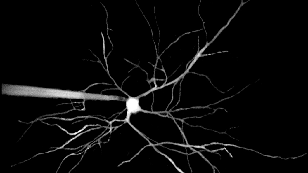
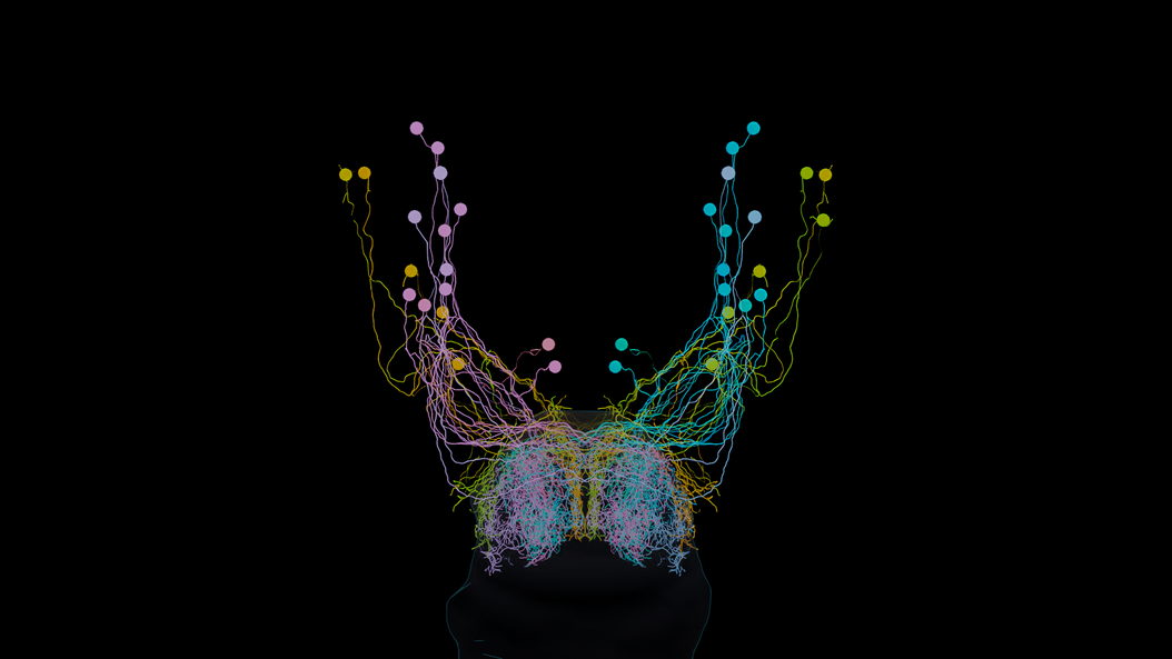
Professor Misgeld is a neurobiologist, who explores the mechanisms that contribute to the degeneration of axons and synapses in neurological diseases and during normal brain development. His research team employs in vivo microscopy in the peripheral and central nervous systems of mice and zebrafish.
Neuroscience
Prof. Dr. Arthur Konnerth
Institute of Neuroscience
Publications
Prof. Konnerth explores basic processes of brain function. By means of electrophysiology, imaging and cell biological approaches, he focuses on synaptic interactions in neuronal circuits in order to achieve a better understanding of mechanisms that underlie learning and memory. A further goal is the elucidation of the neuronal defects that are associated with Alzheimer’s Disease.
Brain Circuit Function & Dysfunction
PI Ruben Portugues
Research Group Brain Circuit Function & Dysfunction
Publications
Having representations of the environment in our brain allows us to interact efficiently with our surroundings, and socially with other individuals. For instance, imagine walking from your house to the train station. Your brain lets you picture the environment and mentally navigate the way. But how is this map encoded in our brain? How does the brain represent the world around it, generate behavior, and adjust to changing conditions? Ruben Portugues, Professor of Brain Circuit Function and Dysfunction, investigates these questions using larval zebrafish as a model organism.
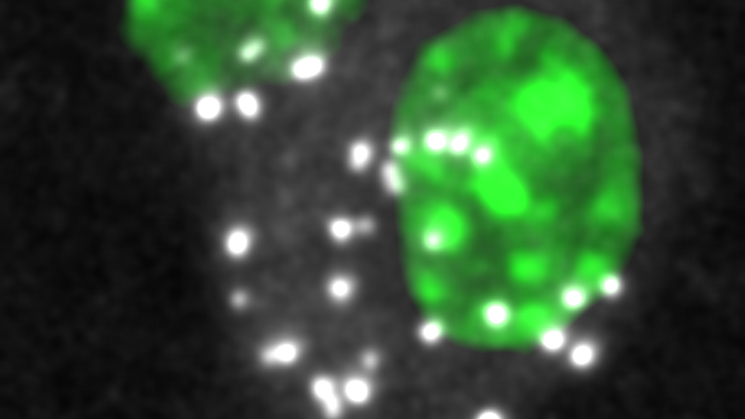
Compared to humans, who have brains with 100 billion neurons, larval zebrafish have small brains with 100 000 neurons. The small size and transparency of these vertebrates make it possible for researchers to monitor their neural activity under the microscope using fluorescent indicators. The researchers can literally see the fish think. To explore how changes in the environment affect neural activity, the researchers create a virtual reality environment with modifiable rules for the fish. They then monitor the fish’s neural activities while they change the rules.
In addition to microscopy, the research team uses a wide variety of methods ranging from in-vivo electrophysiology and optogenetics to large data analysis in order to investigate fundamental biological and neurophysiological questions.
Phase-Contrast X-Ray Imaging
PI Franz Pfeiffer
Chair of Biomedical Physics
Publications
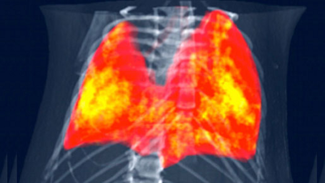
The basic physics principles of x-ray image formation in radiology have remained essentially unchanged since Röntgen first discovered x-rays over a hundred years ago. The conventional approach relies on x-ray attenuation as the sole source of contrast and ignores another, complementary source of contrast. Phase-contrast imaging techniques, on the other hand, offer ways to augment or complement standard attenuation contrast by incorporating phase information. In the recent past several developments have been made that now allow translating x-ray phase-contrast to future clinical applications in radiography and computed tomography.
Biomedical Imaging Physics
PI Julia Herzen
Research Group Biomedical Imaging Physics
Publications
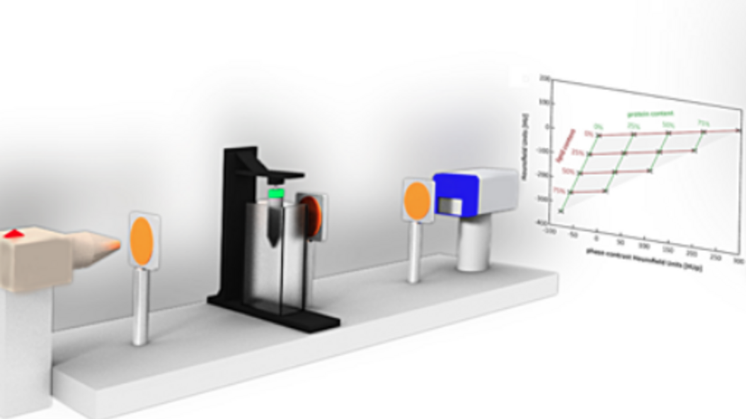
The team around Professor Herzen develops novel X-ray imaging methods using highly brilliant synchrotron radiation and conventional laboratory X-ray sources. They mainly focus on quantitative multi-modal approaches combining spectral and phase-contrast imaging. Currently, they are aiming at applying these methods for improved breast cancer detection and for quantitative 3D virtual histology of human tissue.
More information:
TEDx Talk (Youtube-Video): Novel X-Ray technology that can revolutionize preventive medicine
Neurobiology
Prof. Dr. Harald Luksch
Chair of Zoology
Publications
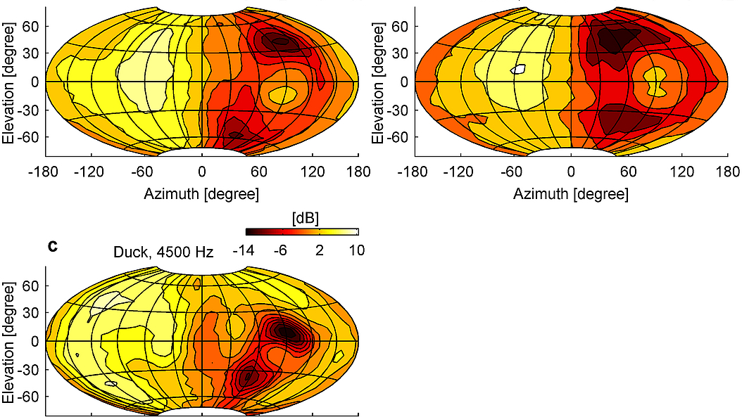
Prof. Luksch has a background in neurobiology and explores sensory processing in the brains of vertebrates. His lab focuses on neuronal networks in the midbrain, a central area for the orientation of the body and sensory organs, e.g., eye movements. His goal is to gain a mechanistic understanding of processes such as object selection and the integration of multimodal stimuli, and to make these findings available as algorithms for technical applications.