Dark-field imaging with x-rays enables the precise diagnosis of respiratory ailments with only very low radiation doses. The method visualizes the scattering of x-rays in tissue using three grating structures and can provide important information on changes in the alveoli.
X-ray images have played a crucial role in medical diagnostics for more than a century – they clearly visualize broken bones and reveal pathological changes in the lung and the female breast. The method is based on a relatively simple principle: The x-ray image shows how strongly the x-rays are attenuated in the tissue, thus producing in essence an x-ray shadow of the tissue.
But the possibilities of classical x-ray imaging are limited: This method can only provide imprecise images of for example structures in soft tissue with weak x-ray absorption properties. And the classic x-ray only captures a part of the changes undergone by the x-rays when they pass through the tissue, meaning that some of the information contained in the rays remains unused.
Scattering visualizes tissue structures
For example, the small portion of the x-ray light which is very slightly deflected from its straight path in the tissue is disregarded. This effect, referred to as scattering, occurs primarily at the interfaces between materials of different densities – for example between air and tissue, such as those found in the alveoli. The x-ray dark-field imaging method, developed by Prof. Franz Pfeiffer, makes it possible to analyze these scattered x-rays; the measurement results provide information about the tissue structures. This means the method can help diagnose changes in the alveoli, thus making an important contribution to the diagnosis of respiratory ailments.
X-rays scatter because they behave as waves. Related visible light phenomena can be found in the everyday world – for example, a rainbow occurs when tiny water droplets in the atmosphere refract white sunlight. Here the intensity of the refraction depends on the wavelength of the light. In the visible spectrum, the wavelength corresponds to the perceived color; thus the rainbow shows the white light split into various different colors.
Scattered light has already been used in investigations with visible light for a very long time. The dark-field microscopy method makes it possible for the microscope to clearly visualize objects which are for the most part transparent. To do so, the object is illuminated with visible light, but only that part of the light which is scattered by the object is considered. As a result, the surroundings of the object where there are no scattering structures appears dark, a factor which gives the method its name.
Grating-based x-ray imaging
However, this kind of microscope cannot be realized using x-rays, because it is not possible to produce the lenses needed for x-rays. Instead of lenses, x-ray dark-field imaging uses gratings as optical elements. These gratings are arrangements of fine lines which alternate between different degrees of x-ray transparency. If light falls on one side of the grating, the light waves which pass through the different openings overlap behind the grating. This overlap – or interference – results in typical patterns of light and dark areas that can be visualized using a detector or photographic film. An object which is in the path of the light changes this pattern; the changes in the pattern make it possible to ascertain the structure of the object.

Download English version of the schematic
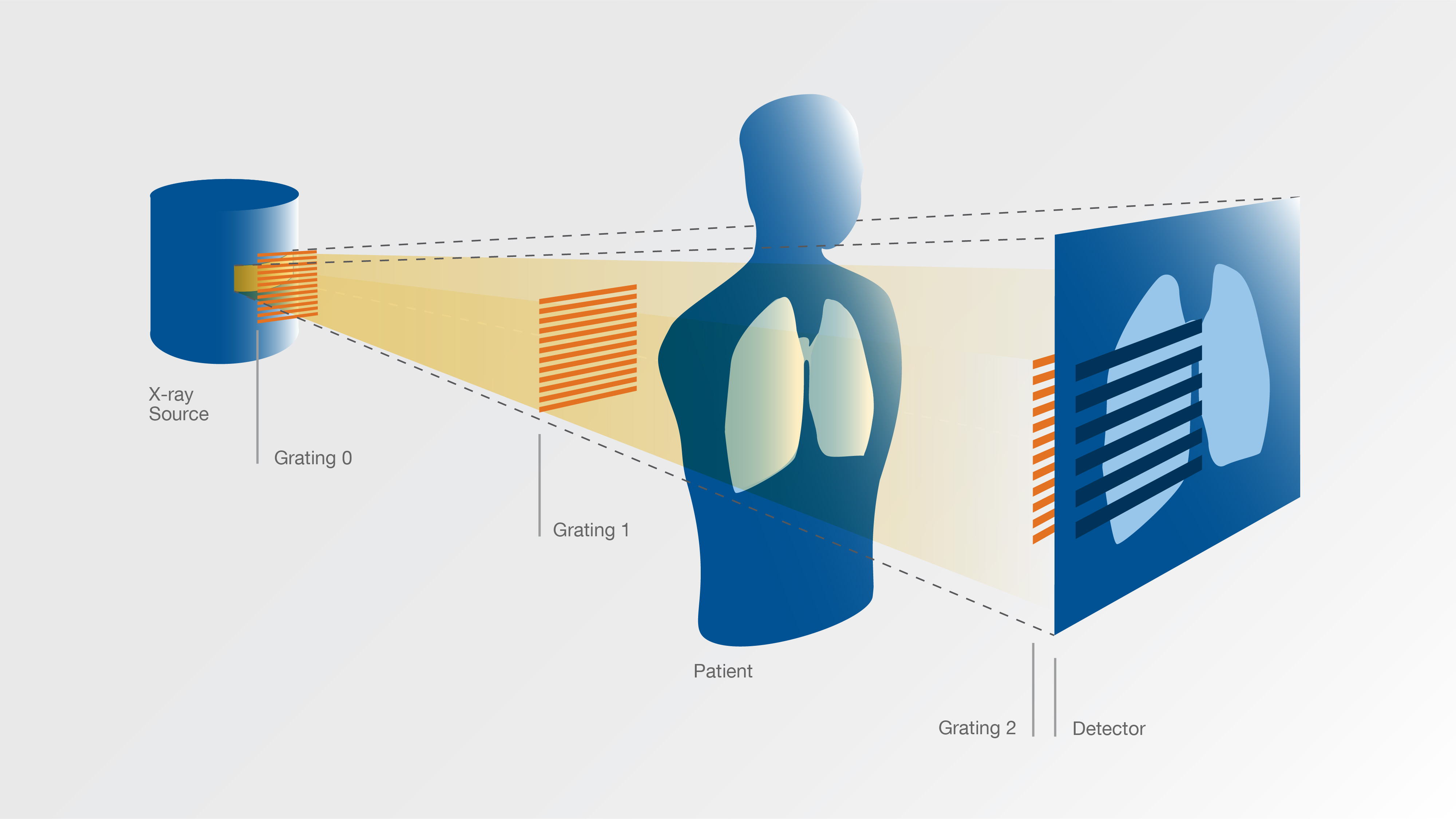
A total of three gratings are used in dark-field imaging with x-rays. The x-rays are produced by a conventional x-ray tube, pass through the three gratings and are then registered by a detector. The detector replaces the x-ray film of the classic x-ray image and works in a manner similar to the chip in a digital camera. Because of the short wavelength of the x-rays, the lines of the gratings are only a few micrometers (thousandths of a millimeter) wide.
During an examination, the patient is located between the second and third grating. The x-ray detector then receives a conventional x-ray image which is overlaid with a pattern of fringes having a width of several millimeters. The scattering weakens this additional pattern so that it is weaker in the parts of the image which visualize areas of the body where much light is diverted by scattering.
In the example of the lung examination, this means that the fringe pattern is the weakest in the areas where regions with intact alveoli are visualized. Specialized software is then used to reconstruct two separate images from this representation – a conventional x-ray image and a dark-field image in which the areas with the intact alveoli appear bright, the areas with damaged alveoli appear dark. The individual alveoli are much too small to be visualized in this type of examination. Nevertheless the method makes it possible to acquire information on their structure.
The three gratings in dark-field imaging
In order to illustrate the roles of the individual gratings, it is best to begin with the second of the three gratings (Grating 1 in the schematic), the phase grating: All of its lines are transparent for the x-rays, but shift their phases to different extents. Behind the grating the various phase-shifted portions of the light generate interference and thus at a certain distance create a create a fine linear periodic pattern with line separations of several micrometers.
If a patient is located in the path of the x-rays between the phase grating and the detector, this line pattern changes due to scattering in the tissue. However, since the lines are too thin to be visualized by a conventional x-ray detector, the third grating (Grating 2) with absorbing lines is positioned shortly before the detector. Via the moiré effect, the interference at this grating turns the fine lines into wider stripes which are then overlaid on the x-ray image.
Coherent light from the x-ray tube
In order for this interference to occur, the x-rays used have to be of a sufficiently high quality – technically, known as "coherent" light. Coherent light can be produced for example by lasers or, for x-rays, by synchrotron light sources – large research facilities which often measure several hundred meters in circumference.
In the dark-field imaging structure the first grating (Grating 0), located immediately in front of the x-ray source, makes it possible to conduct the dark-field measurements using x-ray tubes of the type found in physician's offices. Referred to as a source grating, it consists of an absorbing material in which several fine parallel slits have been made. The x-rays which have left the tube and pass through one of these slits are sufficiently coherent for the purposes of the examination. Thus, the source grating functions like a series of sources of coherent x-rays.
Scientific Contact
Prof. Dr. Franz Pfeiffer
Technische Universität München
Lehrstuhl für Biomedizinische Physik
Munich Institute of Biomedical Engineering
Tel: +49 89 289 12551
E-Mail: franz.pfeiffer@tum.de